[Frontier Science] A New Eye of Biological Detection-Terahertz Imaging
Original BSC China biophysics society
Author | Wang Huabin Yang Zhongbo Chen Ligang
✦
There is a frequency between infrared and microwave in the electromagnetic spectrum ranging from 0.1 THz to 10 THz (1 THz= 1×10⊃1; ⊃2; Hz) electromagnetic wave, called terahertz wave (THz, Figure 1), with a wavelength of 30 μm to 3 mm, is an invisible light wave.
With the rapid development of modern science and technology, terahertz waves have gradually entered people’s field of vision and life. For example, terahertz waves are used in 6G communication. Interestingly, terahertz waves are used in many fields, especially in biomedicine. Biological molecules and water molecules have characteristic absorption in terahertz wave band, and biological samples can be imaged and detected by terahertz wave technology [1-2]. Different from the common biological fluorescence imaging technology, in terahertz biological imaging technology, there is no need to label the sample (such as introducing dye molecules or fluorescent groups) and pretreat it (such as embedding or dehydration), and terahertz photons do little damage to biological samples [3-5]. Terahertz biological imaging has attracted more and more attention of scientists because of its advantages of no marking, no damage and safety, and it has become a new "eye", which is expected to bring new technological revolution to precision medicine. Next, let’s take a look at three different terahertz biological imaging technologies from the perspective of spatial resolution.
✦
✦
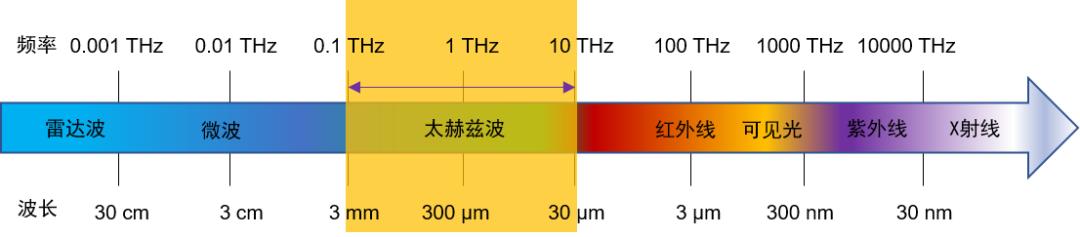
Figure 1. Schematic diagram of the position of terahertz wave in the electromagnetic spectrum.
Sub-millimeter resolution terahertz biological imaging technology
Focusing the terahertz beam emitted from the Emitter on the surface of the sample, and then using the Detector to receive the terahertz light transmitted through or reflected from the surface of the sample, the terahertz imaging of the sample can be realized (Figure 2). This terahertz imaging technology belongs to the traditional far-field detection technology. Due to the limitation of optical diffraction limit, the spatial resolution is generally sub-millimeter, so it is mainly used for biological tissue detection. Using this technology, it is convenient to image and detect diseased biological tissues, such as melanoma tissues (Figure 3).
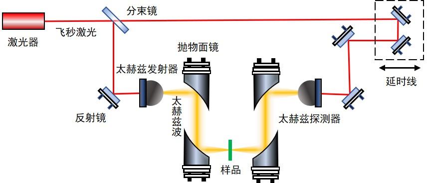
Figure 2. Schematic diagram of submillimeter resolution terahertz imaging technology.
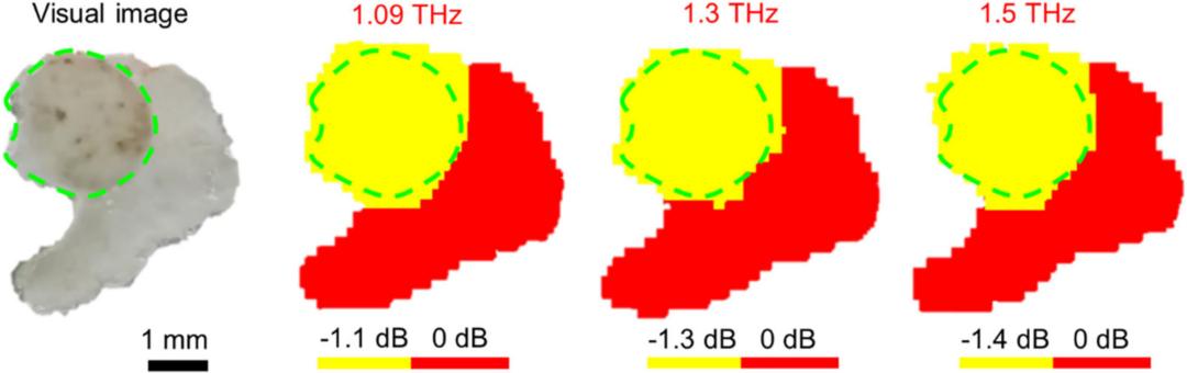
Fig. 3. THz imaging detection of biological tissue [6]. Optical image of mouse skin melanoma tissue (left) and terahertz images at different frequencies. The green circle shows melanoma, and the others are normal tissues.
Microresolution terahertz biological imaging technology
People soon discovered the shortcomings of traditional terahertz imaging technology in resolution, so they developed a new high-resolution terahertz imaging technology [7]. This technology mainly uses a miniature photoconductive antenna detector to detect terahertz waves passing through the sample within a range of several microns from the surface of the sample. Because the distance between the detector and the sample is less than half a wavelength of terahertz wave, the detection belongs to near-field optical technology, which can overcome the limitation of optical diffraction limit and realize micron-scale resolution detection (Figure 4). Using this technology, the researchers monitored the dehydration process of cells by imaging (Figure 5), and they could clearly observe the volume shrinkage of cells and the changes of internal substance concentration during dehydration (arrow).
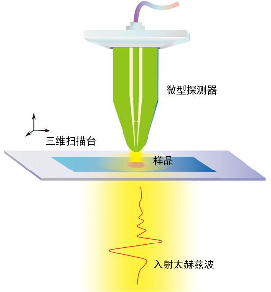
Fig. 4. Schematic diagram of micron resolution terahertz imaging.
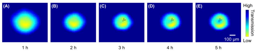
Fig. 5. Terahertz image of cells during dehydration [7].
Nano-resolution terahertz biological imaging technology
Although micron-resolution terahertz imaging technology can carry out biological detection at the cellular level, it cannot carry out imaging research with higher spatial resolution requirements. For example, imaging biomolecules requires terahertz imaging technology with nanometer resolution. In order to solve this problem, scientists combined terahertz technology with scanning nano-probe imaging technology and invented terahertz imaging technology with nano-resolution. In this technology, the terahertz beam is irradiated on the end of the metallic nano-probe, and the terahertz beam is focused to the nanometer scale, thus achieving the purpose of spatial resolution at the nanometer level. On this basis, by analyzing the detected reflected terahertz light, the nano-resolution terahertz imaging of the sample can be finally realized (Figure 6). Using this technology, the researchers successfully realized the terahertz imaging detection of a single protein molecule and obtained its terahertz spectral information (Figure 7).
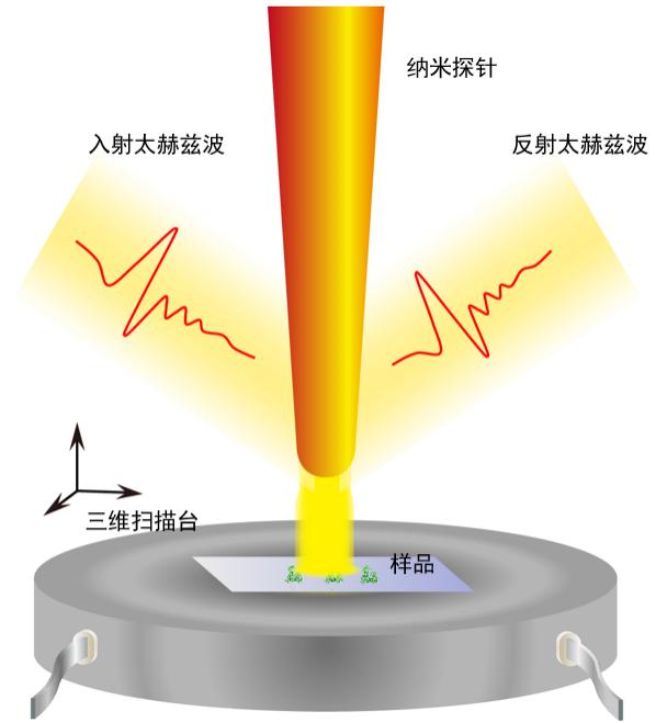
Figure 6. Schematic diagram of nano-resolution terahertz imaging.
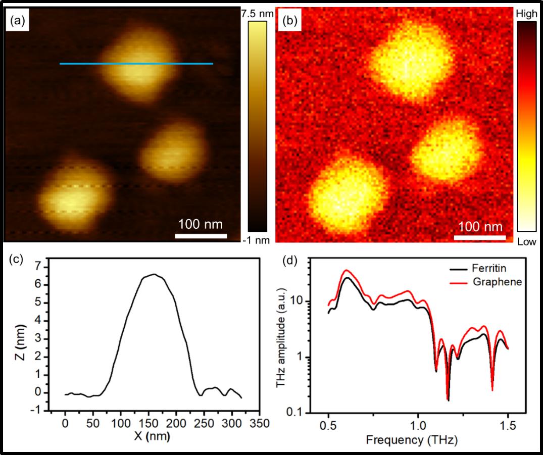
Fig. 7. Near-field terahertz detection of ferritin molecules [8]. (a) Morphology image of atomic force microscope; (b) terahertz images; (c) Height profile (along the blue line in figure (a)); (d) Terahertz frequency domain signals of ferritin molecules and graphene substrates.
Prospect and prospect
Terahertz biological imaging technology is a new frontier cross technology, which can be used to carry out detection and research at three different levels: tissue, cell and biomacromolecule, so it is expected to play an important role in biomedical field. We need to note that the development and application of terahertz biological imaging technology depends on the development of terahertz sources, terahertz detectors and data analysis to a great extent, so the full play of the role and function of this "eye" requires the joint efforts of many disciplines.
references
[1] Yang X, Zhao X, Yang K, et al. Biomedical applications of terahertz spectroscopy and imaging. Trends in Biotechnology 2016, 34(10): 810-824.
[2] Mittleman DM. Twenty years of terahertz imaging invited. Optics Express 2018, 26(8): 9417-9431.
[3] Zhang XC, Shkurinov A, Zhang Y. Extreme terahertz science. Nature Photonics 2017, 11(1): 16-18.
[4] Weissleder R, Pittet MJ. Imaging in the era of molecular oncology. Nature 2008, 452(7187): 580-589.
[5] de Jong M, Essers J, van Weerden WM. Imaging preclinical tumour models: improving translational power. Nature Reviews Cancer 2014, 14(7): 481-493.
[6] Li DD, Yang ZB, Fu AL, et al. Detecting melanoma with a terahertz spectroscopy imaging technique. Spectrochimica Acta Part A: Molecular and Biomolecular Spectroscopy 2020, 234: 118229.
[7] Li ZX, Yan SH, Zang ZZ, et al. Single cell imaging with near‐field terahertz scanning microscopy. Cell Proliferation 2020, 53(4): e12788.
[8] Yang ZB, Tang DY, Hu J, et al. Near-field nanoscopic terahertz imaging of single proteins. Small 2021, 17(3): 2005814.
Brief introduction of the author

Wang Huabin is a researcher at Chongqing Institute of Green Intelligent Technology, China Academy of Sciences, and a member of terahertz biophysics branch of China biophysics society. Main research interests: development and application of terahertz spectroscopy and imaging system.
Lose 12

Yang Zhongbo, assistant researcher of Chongqing Institute of Green Intelligent Technology, China Academy of Sciences, member of terahertz biophysics branch of China biophysics society. Main research direction: terahertz super-resolution biological imaging detection research.
Lose 12

Chen Ligang, assistant researcher of Chongqing Institute of Green Intelligent Technology, China Academy of Sciences, member of terahertz biophysics branch of China biophysics society. Main research direction: terahertz super-resolution biological imaging detection research.
Lose 12
Original title: THz Imaging, a New Eye of Biological Detection [Frontier Science Popularization]
Read the original text